- Quick navigation
- Home
- Open menu
- Page content
- Customer service
- Search
- Footer
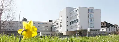
Radiology in Rheinfelden
Radiologie Zentrum Fricktal RZF AG

Radiologie Zentrum Fricktal RZF AG
Radiology in Rheinfelden
Gallery (13)
- Monday8:00 to 18:00
- Tuesday8:00 to 18:00
- Wednesday8:00 to 18:00
- Thursday8:00 to 18:00
- Friday8:00 to 18:00
- SaturdayClosed
- SundayClosed
- Monday8:00 to 18:00
- Tuesday8:00 to 18:00
- Wednesday8:00 to 18:00
- Thursday8:00 to 18:00
- Friday8:00 to 18:00
- SaturdayClosed
- SundayClosed
- Monday
Radiologie Zentrum Fricktal RZF AG – Contacts & Location
Description
The Radiology Center Fricktal in Rheinfelden is a service provider for medical imaging under the direction of Dr. med. Caroline Zähringer. Since 2003, our institute has been the first port of call for radiological examinations in Rheinfelden and the Fricktal region, offering magnetic resonance imaging, computed tomography, mammography, ultrasound, X-rays and bone density measurements.
We are pleased to contribute to precise diagnostics with state-of-the-art technology. As a partner to doctors and patients, we focus on expertise, quality and individual care. Our primary goal is to combine this expertise with the satisfaction of our patients.
The range of services includes- Magnetic resonance imaging 3 Tesla
- Computed tomography 128 sectional images
- Mammography
- X-ray and fluoroscopy
- Ultrasound
- Osteodensitometry (DEXA)
- RIS and PACS (electronic recording of patient data and images)
The Radiology Center Fricktal offers
- Competent care
- Examinations on the latest generation of equipment
- Examination appointments within 24 hours
- Reporting within 24 hours
- Images on CD and email link immediately after the examination
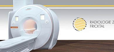
Magnetic resonance imaging 3 Tesla (MRI)
Magnetic resonance imaging is a simple method of looking inside the body. No X-rays are used to generate the images, instead a magnetic field and radio waves are used.
MRI produces high-resolution images of the inside of the body, which can be used to differentiate between healthy and diseased tissue. The stronger the magnetic field, the higher the image resolution and the shorter the examination time. Both devices at the Fricktal Radiology Center have a magnetic field strength of 3 Tesla, making them stronger than conventional devices (1.5 Tesla).Soft tissues are particularly suitable for magnetic resonance imaging; the following organs are therefore examined using MR:
- Brain and spinal cord
- Intervertebral discs
- Joint structures such as cartilage, ligaments and tendons
- Abdominal organs such as bile ducts, small intestine
- Ovaries, uterus, prostate, rectum and pelvic floor
- Vessels
The lack of radiation exposure also makes it suitable for use in children and pregnant women
.Restrictions apply to pacemakers (these patients cannot be examined) and certain heart valve prostheses and aneurysm clips. There are no restrictions for joint prostheses and oesteosynthesis material, such as spinal screws.
Computed tomography with the latest software to reduce the X-ray dose
Computed tomography is an X-ray procedure in which a cross-sectional image is calculated from a large number of individual images
which are taken in a ring around the body. At the beginning of the CT era in the 1970s, the images were very grainy and took minutes to produce. Today, it is possible to create cross-sectional images with a thickness of less than 1 mm and to examine the entire body in just a few seconds.
The CT at the Radiologie Zentrum Fricktal captures 128 cross-sectional images in a single rotation lasting just 0.4 seconds, covering a length of 4 cm in this short time.
Thanks to the very precise images, reconstructions are possible in all planes and in three dimensions, which makes assessment much easier.
Computer technology developed in recent years has made it possible to significantly reduce the radiation exposure of the CT, up to 80% depending on the region being examined! This technology can only be installed on so-called "high-end" devices, such as the one at Radiologie Zentrum Fricktal, and has been used by us since 2012.
The areas of application of CT are:
- Examinations of the chest and abdomen
- of the skeleton
- of the urogenital tract, especially for questions about kidney and ureteral concrements
- the vascular system, in particular for questions about pulmonary embolisms
- the coronary arteries
Mammography
Mammography (MG) is a special X-ray procedure for examining the mammary gland. Using particularly "soft" radiation, the mammary gland tissue is visualized and pathological changes, primarily breast cancer, can be diagnosed.
Our device is completely digital with better image quality and a lower radiation dose compared to conventional devices.
In 1895, Konrad Röntgen discovered X-rays, a short-wave electromagnetic radiation that penetrates tissue and blackens a film behind the tissue to a greater or lesser extent, depending on its absorption.
Today, we still use X-rays, but the film has been replaced by digital detection. This has significantly improved the image quality and reduced the radiation dose.
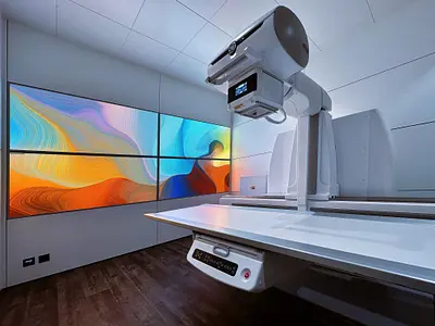
The X-ray machine at Radiologie Zentrum Fricktal has a number of special features. It can take both X-rays and fluoroscopic images (images of motion sequences), it is fully digital and special images such as whole-leg or whole-spine images can be taken.
Ultrasound works with sound waves, without X-rays. The method is comparable to an echo sounder. Sound waves travel from the ultrasound probe into the tissue, where they are reflected to varying degrees depending on the type of tissue and sent back to the probe. The resulting image provides information about internal organs. The sound is reflected from bones and air - as is often present in the gastrointestinal tract - without any information.
Osteodensitometry (DEXA)Bone densitometry uses very weak X-rays to measure the density or calcium salt content of the bone. People with low bone density have an increased risk of fractures. If the bone density is slightly reduced, this is called osteopenia; if it is severely reduced, it is called osteoporosis. 30% of all women develop osteoporosis after the menopause. Medications such as corticosteroids also promote the loss of bone substance.
You can find more information on our website www.radiologiefricktal.ch
- Croatian,English,French,German,Italian,Turkish
- Accepts new patients,Telephone consultation
- By telephone,Free consulting,In-store
- Close to public transport,Close to train station,In city center,Parking site,Wheelchair-accessible,Wheelchair-accessible parking
- Individual practice
- Bill,Cash,EUR
- Doctor, both available
- Categories
- RadiologyRadiological instituteDoctors